
Access for all – No constraints – Radically simplified workflows
Meet Mica at Leica Virtual Campus (live demos, materials and recordings)
Access for all
With Mica YOU save time by eliminating over 85 % of tedious setup steps that require special expertise. With Mica you use 1/3 less time to to the imaging result with half of the training time!
Mica is so easy to use that high-end microscopy from sample to ready analysis results is now in everyone’s reach! All researchers can leverage their microscopy to a new level!
- Intelligent automation
- Intelligent imaging
No constraints
4x more data with 100% correlation
You can image 4 fluorescence channels simultaneously and fast with Leica Mica. Mica combines both widefield and confocal (true point-scanning confocal) microscopy, as well as, Leica developed THUNDER (widefield opto-digital clearing) and LIGHTNING (adaptive deconvolution). You can switch the system from widefield to confocal by press of button: from fast overviews to sharp details in an instant! Last but not least, Mica is also an incubator!
Absolute spatiotemporal correlation
- 4 labels simultaneously
- 4 labels 100% correlated
- Patented FluoSync technology
Seamlessly move from fast overview to high resolution
- Unified imaging modalities (Widefield & THUNDER, Confocal & LIGHTNING)
Point Scanning Confocal - Fully integrated and seamlessly designed incubation
Radically simplified workflows
With Mica’s intelligent automation, you can radically reduce the time and effort from sample to insight by simplifying the entire workflow:
- Sample Finder: searches the sample automatically
- One touch auto illumination: adjusts lighting automatically
- AI-based image analysis: automatically and seamlessly in the workflow (Pixel Classifier, GUI operated annotations, Reusable AI models and project parameters)
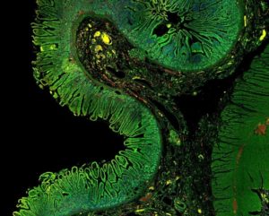
Intestine tissue section acquired with different objectives ranging from low to high magnification (1.6x, 10x, 20x, 63x), using widefield and confocal imaging. 20x widefield images are processed with THUNDER and 63x confocal images with LIGHTNING. Nuclei are labeled in blue, mitochondria in green, and detyrosinated tubulin in red.
Leica Virtual Campus
You can learn more about Mica also from the Leica Virtual Campus – live demos, materials and recordings.