Hämeenlinna

Kaivokatu 16
13100 Hämeenlinna
Y-tunnus: 0499964-0

Phone +358 3 615 370

Espoo

Sinimäentie 10 C
02630 Espoo

Oulu – Technical Service

Aapistie 7 B
90220 Oulu

Orders, product search/enquiries and other questions: info(@)immunodiagnostic.fi

Contact us Technical support and service
Move to content

Fluorescence

Insights into the structures and dynamics of life

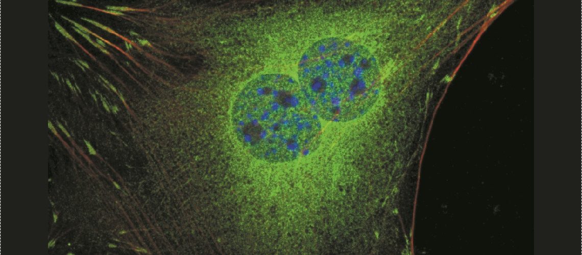
Fluorescence microscopy is essential technique for studying biological processes. Fluorescence can be used to visualize specific subcellular structures in fixed cells, and reveal connections between dynamic processes in live cells and tissues. Without fluorescence labeling, few cellular process can be visualized since most biological samples are almost transparent. A broad range of advanced Live-cell Imaging Techniques can be employed by researchers to probe the complexities of cellular processes. Fluorescence imaging is central to many of these imaging techniques.

Sensitivity,quality and speed

To obtain excellent fluorescence images you need a highly sensitive camera delivering a high signal-to-noise ratio and a large dynamic range resulting in a crisp fluorescence signal. Live-cell imaging often requires a high acquisition speed to capture fast dynamic processes.

Leica DFC3000G
1.3 MP monochrome camera for basic fluorescence applications such as documentation of fixed, immunostained samples

Leica DFC7000T
2.8 MP cooled color camera for brightfield and fluorescence documentation of fixed cells and tissues

Leica DFC9000
4.2 MP cooled monochrome fluorescence scmos camera for advanced applications like high-speed live cell imaging, FRAP, and ratio measurement

Contact us for more information!

Contact us

Markku Saari

Product Manager, Microscopy and Imaging

Vesa Ylöstalo

Product Manager, Microscopy and Imaging

Toni Vormisto

Product Manager, Stereo and material microscopes

Janne Ylärinne, Ph.D.

Sales Director