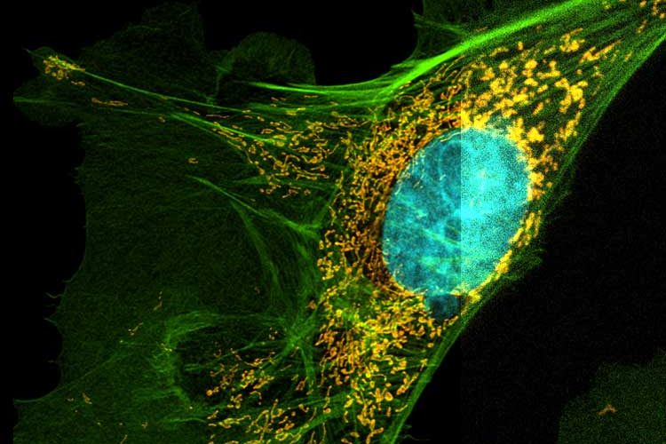
Confocal Microscopes
Confocal microscopes from Leica Microsystems are partners in top level biomedical research and surface analysis in material science applications, offering unprecedented precision in three-dimensional imaging and exact examination of subcellular structures and dynamic processes.
High-speed imaging supplies the data for a wide range of integrated analytical techniques. Our confocal microscopes are based on a modular concept that enables flexible upgrading and integration of innovative technology all the way to the nano range with STED 3X.
Super-Resolution Microscopes and Nanoscopes
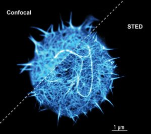
Super-resolution microscopes and nanoscopes overcome the diffraction limit of light and allow investigators to study subcellular structures in greater detail than achieved with a standard confocal microscope. With the possibility of resolutions down to 30 nm with STED (Nobel Prize in Chemistry in 2014) as well as sub-cellular dynamics can be studied at the nanoscale. Sub-diffraction colocalization analysis reveals interactions unprecedented detail. Enabling novel discoveries to be made in the fields of virology, immunology, neuroscience and cancer, super-resolution is on its way to becoming the new gold standard in light microscopy.
Your entry has never been easier than today with HyVolution 2 and STED 3X.